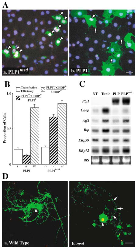
Figure 1. The UPR Is Induced in Cells Expressing Mutant PLP1.
(A) Transfected COS-7 cells expressing mutant (Aa) but not wild-type (Ab) PLP1 (green) localize CHOP (red) in the nucleus (blue). Arrowheads show CHOP+ cells (pink/white nuclei). Scale bar, 20 μm.
(B) Morphometry of transfected COS-7 cells showing transfection efficiency (bars i and iv) and PLP1+/CHOP+ cells expressed as a proportion of transfected (ii and v ) or stressed (iii and vi) cells.
(C) Northern blots show induction of UPR effector genes in 293T cells untreated (NT), treated with tunicamycin (Tunic), or transfected with wild-type or mutant PLP1.
(D) Oligodendrocyte progenitors from wild-type mice (Da) elaborate cell processes, differentiate, and incorporate Plp1 gene products into the plasma membrane (green). Progenitors from msd mice also extend processes, which are subsequently resorbed (arrows) after the cells differentiate and express the Plp1 gene (Db). These cells localize CHOP (red) in the nucleus which appears as yellow/orange (arrowheads). Scale bar, 10 μm.

