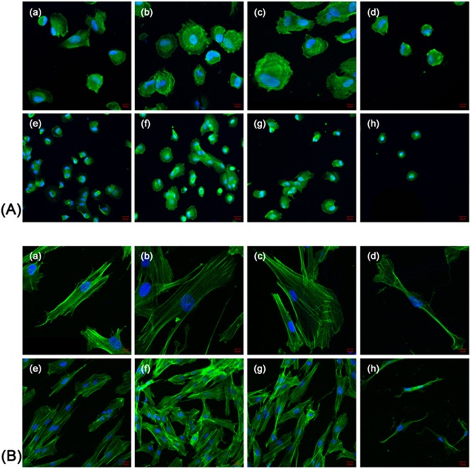
Fig 7. Confocal laser scanning microscopy observations of the HGFs on the control and plasma treatment surfaces.
Confocal laser scanning microscopy images of HGFs on zirconia disks at 3 h (A) and 24 h (B) of culture. The helium plasma treatment time are 30 s (a, e), 60 s (b, f), 90 s (c, g), and 0 s as controls (d, h). High magnification (a-d), scale bar = 10 μm. Low magnification (e-h), scale bar = 20 μm.

