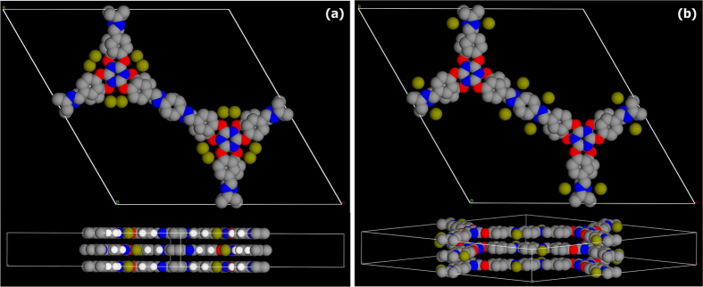
Figure 5. A DMol3 (DFT-D corrected) energy and geometry optimization of the Pd-COF interactions. Several starting models with different Pd position were attempted and it yielded two low energy configurations.

(a) In the minimized geometry, the Pd atoms resided in the small clefts formed around the triazine core lined by the ether and the N and C of the triazine rings. A b-axis view showed that they were lined well with the layer. (b) Another minimized configuration included the interaction of the Pd atoms with the nitrogens of the Schiff bond. It can be seen that the Pd atoms align with the N atoms. In both cases the unit cell was retained and framework atoms were frozen in P6/mcc configuration. Color code: Pd- olive green; O- red; N- blue; C- grey and H- white.
