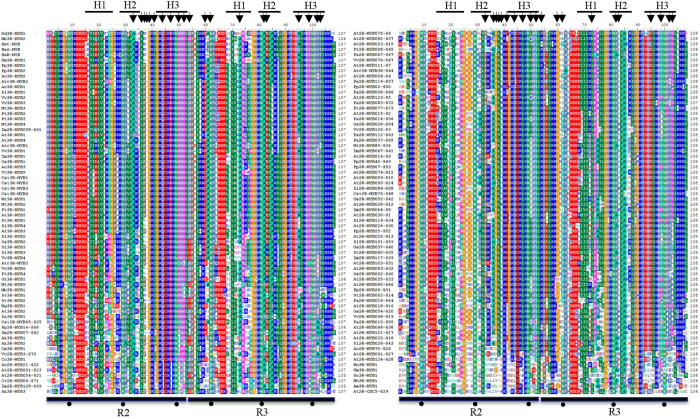
Figure 6. Multiple alignments of R2R3 MYB domains of the representatives of 73 2R-MYB subfamilies and 63 eukaryotes 3R-MYBs.
The three α-helices in each repeat are indicated by a black bold line and marked as H1 to H3. The black arrows indicate the conserved residue changes between the typical plant 2R-MYBs and the early-deriving plant 2R-MYBs and eukaryote 3R-MYBs. The asterisks indicate the three highly conserved tryptophan residues (W) in each MYB repeat.

