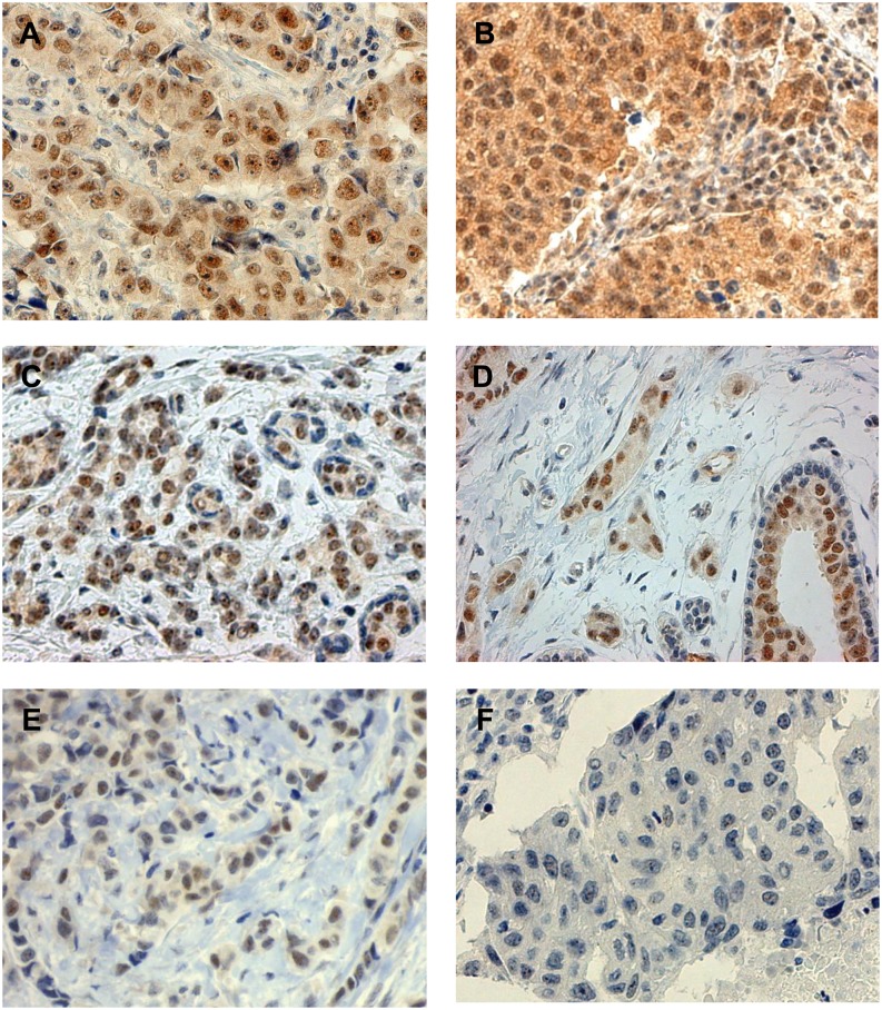
Fig 1. Immunohistochemical staining patterns of WRAP53 in breast cancer.
A. and B. Positive nucleus/positive cytoplasma, C. and D. positive nucleus/negative cytoplasma (with normal mammary epithelial cells), E. positive nucleus/negative cytoplasm (invasive lobular carcinoma), and F. negative nucleus/negative cytoplasm.

