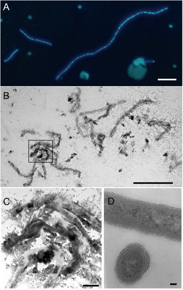
Fig. 6.

Bacterial shape of A. longa SW024T. Fluorescence microscopy of A. longa SW024T stained with DAPI, bar = 5 μm (a). Transmission electron microscopy of A. longa SW024T culturing in MB medium without staining, bar = 10 μm (b) and magnification of aggregate section in the boxed area, bar = 1 μm (c). Transmission electron microscopy of A. longa SW024T using ultramicrotomy, including transections and longitudinal section, bar = 50 nm (d)
