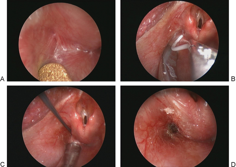
Fig. 3.

Direct laryngoscopy. (A) Endoscopic view of the left pyriform sinus. Note the small mucosal-lined opening at the lateral aspect of the sinus. (B) Placement of a small catheter endoscopically, resulting in immediate deflation of the mass and verifying the diagnosis. (C) Electrocautery catheter device being inserted into the tract. (D) Postoperative view of the cauterized sinus tract.
