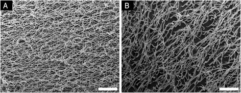
Fig. 2.

Scanning electron micrographs of two small fiber models taken parallel to the substrate at the same magnification. a The 2 × 2 pattern has the smaller diameter fibers and the highest density of fibers at 25 per 100 μm2. b The 5 × 5 mold has larger diameter fibers and a density of 4 fibers per 100 μm2. Scale bars are 20 μm
