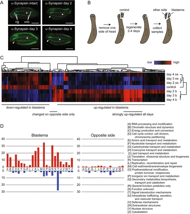
Fig. 1.

Identification of genes differentially expressed during head repair and regeneration. a Half-head regeneration. Anti-Synapsin antibody was used to label the central nervous system of intact animals and animals that had regenerated for two to 4 days following half-head amputation. Samples were imaged from the ventral side with anterior to the left, and the amputated portion of the head in regenerates is toward the bottom. cg = cephalic ganglia; vnc = ventral nerve cords. Asterisks mark the pharynx, and triangles point to where the nervous system is regenerating in the blastema. Scale bar = 0.5 mm. b Sample collection strategy. One side of the head was removed from intact animals and saved as the non-regenerating control sample. After two to four days of regeneration, samples were collected from the blastema and non-regenerating opposite side of the head. c Heat map summary of changes in gene expression in the blastema and other side of the head during a time course of half-head regeneration. Low expression is represented in blue and high in red. Control = non-regenerating samples. Day 2 b, day 3 b, and day 4 b = blastema samples collected on day two, three or four of regeneration. Day 2 os, day 3 os, and day 4 os = samples collected from the opposite side of the head on day two, three, or four of regeneration. d Categorization of differentially expressed genes based on the function of their homologs in other species using Clusters of Orthologous Groups. Red bars show the number of upregulated genes in each functional group in either the blastema or opposite side tissue relative to control. Blue bars represent downregulated genes
