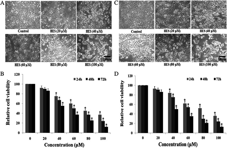
Fig. 1.

Hesperidin (HES)-induced morphological change and anti-proliferation in HeLa cells and HT-29 cells. a and c The morphology of the HeLa cells and HT-29 cellswas examined using a phase contrast microscope after treatment with HES. After treatment with HES (0, 20, 40, 60, 80, and 100 μM) for 48 h, Cells showed numerous morphological changes. Scale bar = 100 μm. b and d HES-induced inhibition of proliferation in HeLa cells and HT-29 cells. Cells were treated with HES at concentrations of 0, 20, 40, 60, 80 and 100 μM for 24, 48, and 72 h. MTT assay results are reported as cell viability (%) relative to the control. All data were normalized to the control group, which was set at 100 %. HES inhibited proliferation of HeLa cells and HT-29 cells in a concentration- and time-dependent manner; *p <0.05 versus control group (0 μM) (two-way ANOVA followed by Tukey’s post hoc test)
