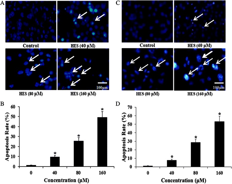
Fig. 2.

HeLa cell and HT-29 cell apoptosis after treatment with hesperidin (HES) observed using Hoechst 33342 staining. a and c HeLa cells and HT-29 cells were treated with HES (0, 40, 80, and 160 μM) for 48 h. Apoptotic cells (Arrows) exhibited morphological changes in the nuclei typical of apoptosis. Scale bar = 100 μm. b and d HES-induced increase of apoptosis in HeLa cells and HT-29 cells. Cells were treated with HES at concentrations of 0, 40, 80 and 160 μM for 48 h. The level of apoptosis was analyzed by Hoechst 33342 staining; *p <0.05 versus control group (0 μM) (one-way ANOVA followed by Tukey’s post hoc test)
