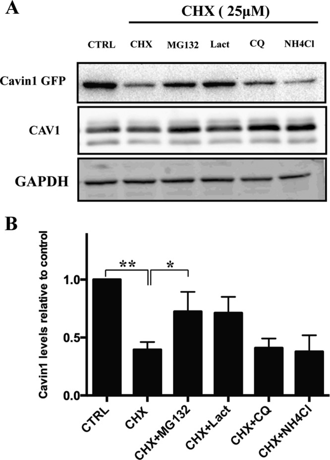
FIGURE 1:

Cavin1 turnover is mediated by the proteasome. (A) PC3 cells overexpressing WT cavin1-GFP were treated with 25 μM CHX in the presence or absence of MG132 (10 μM), Lact (10 μM), CQ (10 μM), or NH4Cl (10 mM) for 6 h and were subsequently immunoblotted for GFP, CAV1, and GAPDH as loading control. (B) Quantification of WT cavin1-GFP levels after inhibitor treatment normalized to untreated samples from three to four independent experiments. Error bars represent SD. *, p < 0.05; **, p < 0.01. Representative uncropped immunoblots are shown in Supplemental Figure S2.
