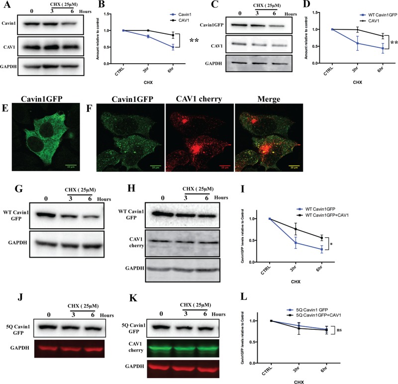
FIGURE 4:
Analysis of cavin1 turnover in model cell lines. (A) A431 cells were treated with 25 μM CHX for the indicated time period following lysis and immunoblotting for cavin1, CAV1, and GAPDH. WT cavin1-GFP was exogenously expressed in PC3 (C), a CHX chase assay was performed as above, and immunoblotted for GFP, CAV1, and GAPDH. Quantification of protein levels by Western blots at various time points is indicated in B and D. Each point represents the mean of three to four independent experiments, and error bars indicate SD. **, p < 0.01. Subcellular distribution of cavin1-GFP (E) upon overexpression in MCF 7 cells and when coexpressed with CAV1-cherry (F). CHX chase assay upon overexpression of WT cavin1-GFP (G) and 5Q cavin1-GFP (J) in MCF7 cells and with coexpression of WT cavin1-GFP (H)/5Q cavin1-GFP (K) and CAV1-cherry. Quantification of total WT cavin1-GFP (I) and 5Q cavin1-GFP (L) levels at each time point normalized to untreated samples from three independent experiments. Data are presented as mean ± SD. *, p < 0.05; ns, no significant difference. Representative uncropped immunoblots are shown in Supplemental Figure S3. Western blot images shown in Figures 2B and 4C (WT cavin1-GFP turnover upon overexpression in PC3 line) originate from different replicates, but quantifications shown in Figures 2E and 4D are the same.

