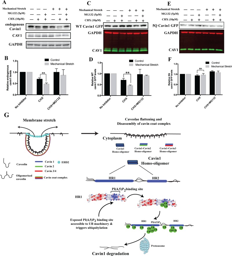
FIGURE 5:
Mechanical stretch stimulates cavin1 turnover. (A) A431 cells expressing WT cavin1-GFP or 5Q cavin1-GFP were subjected to cyclical stretch or rest (control) in the presence or absence of specific inhibitors for 6 h (described in Materials and Methods). Subsequently cells were lysed in lysis buffer A and immunoblotted for endogenous cavin1, cavin1-GFP CAV1, and GAPDH. Quantification of total endogenous cavin1 (B), WT cavin1-GFP (D), and 5Q cavin1-GFP (F) levels was done by normalizing inhibitor-treated samples to untreated samples in respective resting or cyclical stretch conditions. Data are presented as mean ± SD from three independent experiments. **, p < 0.01; ns, no significant difference. Immunoblots for WT cavin1-GFP (C) and 5Q cavin1-GFP (E) levels upon cyclical stretch. Representative immunoblots for endogenous cavin1 and quantification of endogenous cavin1 protein levels from three independent experiments (see panel B) are shown from A431 cells transfected with WT cavin1-GFP. Representative uncropped immunoblots are shown in Supplemental Figure S3. (G) Model of caveolae disassembly and fate of cavins after release into the cytosol.

