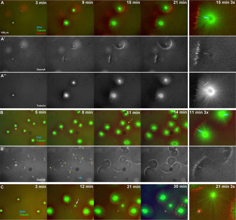
FIGURE 7:
Chromatin spatially biases CPC recruitment to monopolar sperm asters in egg extract. Permeabilized sperm was added to interphase egg extract supplemented with probes for DNA, tubulin, and the CPC (GFP-DasraA). (A–C) Image sequences chosen to emphasize different aspects of this reaction. The last in each series is a 3× magnification. Time points are as indicated. (A) Two isolated sperm asters. Note polarized microtubule distribution and formation of an arc of CPC-positive bundles on the periphery of each aster on the same side as the DNA. (A′) Only the DasraA channel for the same images as A. Note that the DasraA probe binds sperm chromatin. (A′′) Only the microtubule channel. (B) Field at higher sperm density. (B′) Same images in DasraA channel alone. Seven of eight of the asters polarize early (yellow p), and one aster never polarizes (cyan np). Later most asters contact a neighbor and form a CPC-positive interaction zone; yellow arrow at 11-min time point (B). (C) Field that includes a centrosome not attached to a sperm nucleus (white arrow in 12-min image). The centrosome nucleated a weaker aster than the sperm-attached centrosomes. The free centrosome aster did not recruit CPC to its periphery until it contacted a neighboring aster (30-min time point). The bright dot on the lower left side of the free centrosome aster is DNA without a centrosome. With regard to A–C, see Supplemental Movies 7A–7C, respectively.

