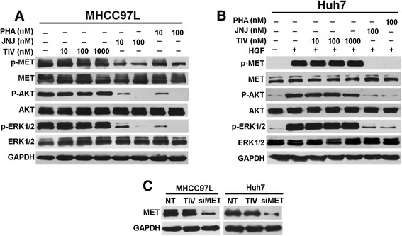
Fig. 2.

Effect of tivantinib on MET signaling in HCC cells. a Western blot analysis was conducted to measure effect of tivantinib on constitutive p-MET and downstream effectors in MHCC97L cells. Cells were treated with the indicated concentrations of tivantinib (TIV), JNJ-38877605 (JNJ) and PHA-665752 (PHA) in DMEM containing 10 % FBS for 4 h before protein extraction. b Western blot analysis was performed to detect effect of tivantinib on HGF-stimulated p-MET and downstream effectors in Huh7 cells. Cells were starved in medium containing 1 % FBS for 12 h before adding the indicated concentrations of compounds. After incubation for 4 h and stimulation with 50 ng/ml HGF for 10 min, cells were lysed for Western blot analysis. (c) MHCC97L and Huh7 cells were treated with 1000 nM tivantinib in DMEM containing 10 % FBS for 24 h before protein extraction. The MET siRNA-transduced cells were used as controls
