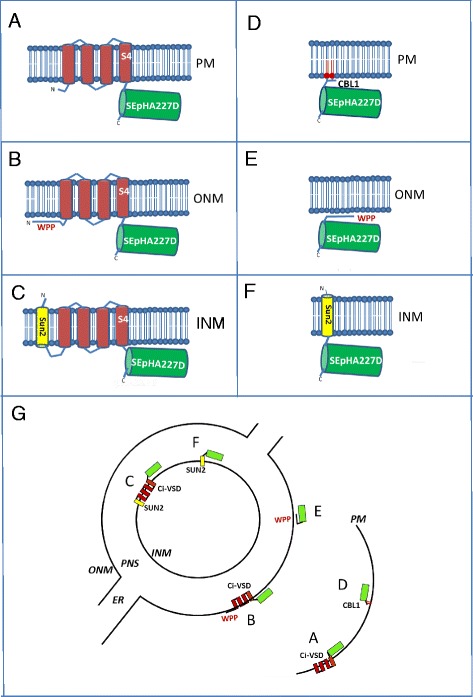
Fig. 1.

Diagrams of GEVIs and derivatives used in this study and predicted membrane localizations. GEVIs include: A ArcLight, which consists of SEpHluorinA227D fused to the Ci-VSD (transmembrane domains indicated as red bars with the voltage-sensing domain in S4); B ArcLight fused at the N-terminus to outer nuclear membrane (ONM)-tethering sequence WPP; C ArcLight fused at the N-terminus to inner nuclear membrane (INM) transmembrane protein SUN2. The derivatives, which do not contain Ci-VSD, include: D SEpHluorinA227D fused to the plasma membrane (PM)-tethering sequence CBL1; E SEpHluorinA227D fused at the N-terminus to WPP; F SEpHluorinA227D fused at the N-terminus to SUN2. Part G shows the predicted membrane localizations of these proteins. The sector letters A-F correspond to the diagram letters. The endoplasmic reticulum (ER) is continuous with perinuclear space (PNS). For simplicity, nuclear pores are not shown. Drawing is not to scale
