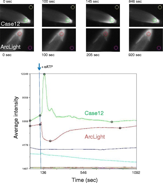
Fig. 4.

Comparison of Case12 and ArcLight responses to eATP. Top: MiCAM images of root tips of plants expressing ArcLight and Case12 with colored circles indicating the root and background regions used for the graphs. The images correspond to the beginning of the experiment (0 s), addition of ATP (100 s), highest response (145 s, Case12, increase of fluorescence; 205 s ArcLight, decrease of fluorescence) and recovery (846 s Case12; 920 s ArcLight), which can also be seen in the open black circles on the traces. Bottom: MiCAM raw data files were imported into Metamorph and combined into one stack for comparison of fluorescence intensity changes. The traces derived from the colored circled areas at the top are displayed over a time period of 1092 s. Either 2 mM ATP or buffer was added at approximately 100 sec as indicated by the blue arrow. The red and green traces represent the responses of ArcLight and Case12, respectively, to eATP addition. Pink and gold traces show the corresponding backgrounds for ArcLight and Case12, respectively. Turquoise and blue traces show the buffer controls for ArcLight and Case12, respectively. Dark red and dark green traces indicate background for buffer controls for ArcLight and Case12, respectively (MiCAM images not shown)
