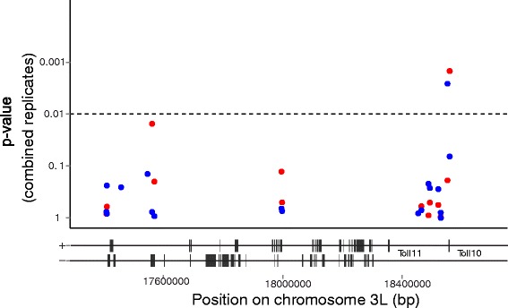
Fig. 3.

Individual genotyping confirms nucleotide variation significantly associated with P. falciparum infection outcome. Fine mapping by logistic regression analysis of Fd03 colony SNPs within locus 3.1 using individual mosquito SNP genotypes. Association is calculated across both biological replicates (the original infection used in pooled sequencing and genotyping of individuals deconvoluted from the pools, and a second independent infection of the same colony). Red points indicate p values for association with the phenotype, oocyst intensity (comparison between individuals from high and low pools), blue points indicate p values for association with oocyst infection prevalence (comparison between individuals from zero and all infected (combined low + high) pools). The dashed line indicates the significance threshold (p = 0.01). Genes within the locus are indicated beneath the plot; genes on the forward (+) and reverse (−) strands are shown separately, positions of TOLL 11 and TOLL 10 genes are indicated
