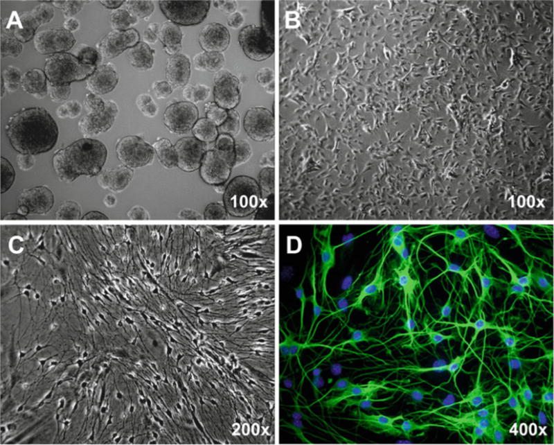
Fig. 2.

Culture and differentiation of enteric neural stem/progenitor cells from 8-week-old mouse. (a) Neurosphere culture for 7 days. (b) Monolayer culture. (c, d) Differentiation for 4 days. Representative phase-contrast (a–c) and immunofluorescent (d) micrographs are shown. Standard immunocytochemistry was performed using rabbit anti-Tuji1 polyclonal antibody (1:5,000, Sigma) and Alexa Fluor® 488 (green) conjugated donkey anti-rabbit secondary antibody (1:400; Invitrogen). Hoechst 33258 was used for counterstaining of nuclei (Blue)
