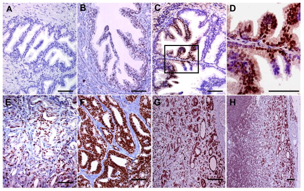
Figure 7. Immunohistochemical staining of prostate tissue and metastases using anti-P-S319-Runx2 antibody.

Representative samples from TMAs are shown. (A) normal prostate tissue, (B) benign prostate hyperplasia, (C) prostate intraepithelial neoplasia (PIN), (D) higher power view of boxed region in C showing strong nuclear staining of basal cells, (E) moderate staining in low Gleason score PCa, (F) very strong staining in high Gleason score PCa, (G) lymph node metastasis. (H) lower power view of lymph node metastasis showing strong staining in extra-capsular spread (ECS). Bars = 100 μm
