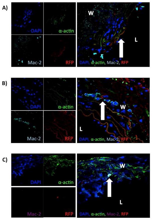
Figure 2.
Confocal microscopy reveals integration of male MSC into aortic wall. A) Convergence of channels of confocal microscopy revealing integration of male MSC into the aortic wall at (arrow) day 3. B) Convergence of channels of confocal microscopy revealing integration of male MSC into the aortic wall (arrow) on day 7. C) Convergence of channels of confocal microscopy revealing integration of male MSC into the aortic wall (arrow) on day 14 (L=lumen, W=wall).

