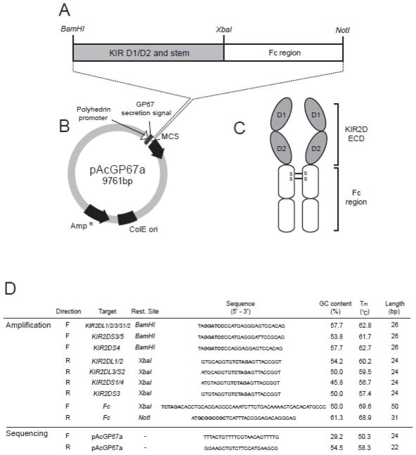
Figure 1.
(A) Schematic diagram showing the configuration of a recombinant KIR-Fc fusion gene. The recombinant fusion gene consisting of the D1, D2 and stem domains of a KIR2D molecule (grey box) and the Fc region of a human IgG1 antibody (white box) is cloned using BamH1 and Not1 restriction sites into the multiple cloning site (MCS) of the pAcGP67a vector (B) in frame behind the GP67 secretion signal sequence. Transfection into insect cells produces a soluble recombinant KIR-Fc dimer (C) consisting of the D1, D2 and stem regions (extracellular domain – ECD) of the KIR molecule (grey ovals) and the Fc region (white ovals) of the IgG1 antibody. (S-S) shows the location of the disulphide bonds that lead to formation of a dimer.
(D) Table listing the primers for amplification and sequencing of KIR-Fc fusion genes representing inhibitory KIR2DL1, 2DL2/3 and activating KIR2DS1, 2DS2, 2DS3, 2DS4 and 2DS5. The properties of each primer, including the GC content, melting temperature (Tm) and length are listed to the right. Restriction sites are shown in bold when present in a primer sequence.

