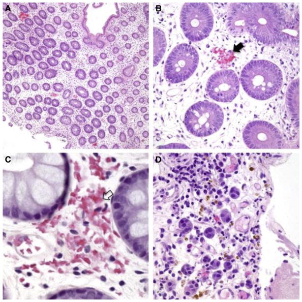
Figure 1.
Histologic samples showing mucosal hemorrhage (H&E, biopsies from 4 iTMA patients, A–C: colon, original magnification ×100, ×400, ×600, respectively, D: stomach, ×400). (A) Patchy distribution of fragmented RBC extravasation located at and/or close to the disrupted capillaries in the lamina propria. (B–C) Capillaries demonstrate fragmented RBC extravasation with partial (B, black arrow) and total (C, clear arrow) disruption of the wall. (D) Hemosiderin deposits (stained brown) and hemosiderin-laden macrophages represent remote mucosal hemorrhage. Nikon Eclipse 80i, 10x/0.30, 40x/0.75, 60x/0.85, Diagnostic Instruments 14.2 Color Mosaic, Spot software.

