Figure 2.

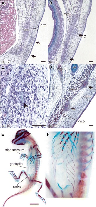
Gastralia of the American alligator ( Alligator mississippiensis ). The embryos were staged according to Ferguson (1985) [26]. (A) Transverse section of the ventral trunk of an embryo at stage 17. Formation of the gastralia begins with condensation of cells (arrows) in the dermis (drm). Alcian-blue, hematoxylin and eosin stains; scale bar, 100 μm. (B) Transverse section of the ventral trunk of an embryo at stage 19. The distance between the primordial gastralia and the rectus abdominis muscle (ram) decreases. Alcian-blue, hematoxylin and eosin stains; scale bar, 100 μm. (C) Enlarged image of the primordial gastralia, showing the matrix that is stained with Alcian blue (arrowhead), which appears transiently before the bony tissue is formed. Alcian-blue, hematoxylin and eosin stains; scale bar, 50 μm. (D) Transverse section of the ventral trunk of an embryo at stage 22. The gastralia contact the rectus abdominis muscle. The ventral cutaneous branch of the intercostal nerve (vcb) runs adjacent to the margin of the gastralium. Alcian-blue, hematoxylin, eosin and immunohistochemistry with anti-acetylated tubulin antibody (T6793, Sigma-Aldrich) stains; scale bar, 100 μm. tvm, transversus ventralis muscle. (E) Ventral view of a stage 25 embryo. Alizarin red and Alcian blue stains; scale bar, 1 cm. (F) Enlarged image of E.
