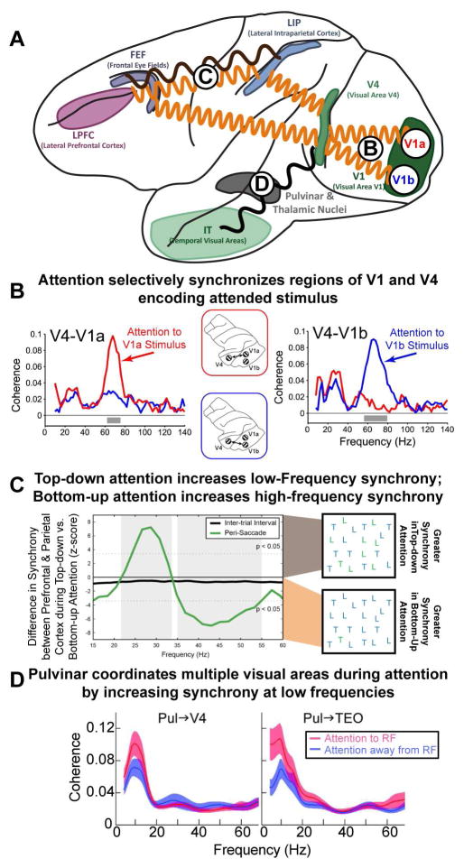
Figure 3. Attention dynamically changes the effective connectivity between brain regions.
(A) Attention modulates the synchrony between different brain regions. A minor subset of the effects of attention is outlined. Circles with letters refer to other parts of the figure. (B) Attentional modulation is specific to selected objects: synchrony between V4 and V1 is specific to those regions that encode the currently attended stimulus (red for a stimulus encoded by V1a; blue for V1b). Note that these changes in the pattern of synchrony overlay the underlying anatomy and can occur rapidly, with each shift in attention. Adapted from (Bosman et al., 2012). (C) There is also flexibility in the frequency of oscillations between brain regions. Internal (top-down) direction of attention and external (bottom-up) capture of attention emphasize different frequency bands between the same brain regions. Synchrony between prefrontal cortex (PFC) and parietal cortex (PPC) changed frequency depending on the type of attention. When attention was externally captured by a salient stimulus, this was reflected in neural activity in PPC first, followed by PFC. In addition, synchrony between PFC and PPC was observed at high-frequencies (~45 Hz; shown as negative deflection). In contrast, when attention was internally directed by the memory of a target stimulus (as in visual search) neural activity was found first in PFC and then PPC and PFC-PPC synchrony was observed at low frequencies (~25 Hz; shown as positive deflection). Adapted from (Buschman and Miller, 2007) (D) Sub-cortical regions, such as the pulvinar, play an important role in attention. In addition, they may act to coordinate activity across cortical regions: attention increased low-frequency synchrony between the pulvinar and V4/TEO (left/right, respectively), when allocated at the receptive field (RF) represented by the recorded neurons (ATT to RF) relative to a different RF location (ATT away from RF). This may organize higher-frequency oscillations, facilitating the establishment of synchrony observed between cortical regions (as in B and C). Adapted from (Saalmann et al., 2012).

