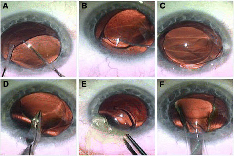
Figure 1.

IOL explantation. Opening of the same corneal incision of the previous cataract surgery and paracentesis, injection of viscoelastic and freeing of the optic of IOL from the capsular bag (A). Dialing of the first haptic to explant it outside the capsular bag (B), and then dialing of the IOL to explant the whole IOL outside the capsular bag (C). Cutting part of the IOL optic with scissors for easy explantation through the small wound (D). Explantation of IOL haptic, then the optic, then the other haptic through the same small wound without widening it (E). Implantation of the 2nd IOL by injector through the main wound (F).
