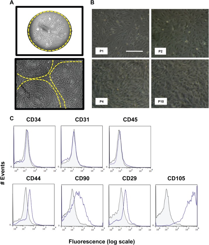
Fig 4. Isolation and characterization of mesenchymal stromal stem cells from murine dermis.
The mesenchymal stem cells from murine dermis were selected for their ability to form colonies at low densities (CFU), as shown in the micrograph (A). These colonies expand and form a confluent layer at P0, as shown in (A). These cells are then passaged whenever they reached 80% confluence and expanded until passage 10 (B). Cells at P4 were used to detect the presence of positive and negative markers of MSCs by flow cytometry, namely: CD34, CD31, CD45, CD44, CD90, CD29 and CD105 (C). All groups marked with conjugated antibody (blue line) were compared to their respective controls (gray filled line); at least 50.000 events were collected for analysis. IgG isotype controls conjugated with Alexa 488 and APC were used as negative controls. Results are representative of those obtained in three independent experiments performed in triplicates.

