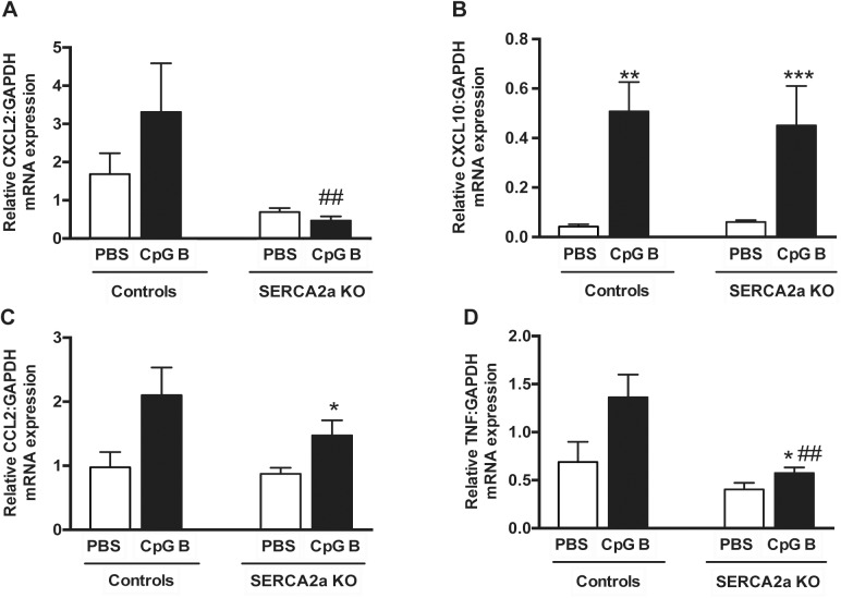
Fig 4. Quantitative PCR on left ventricle myocardial tissue of mice with HF 8 weeks after SERCA2a gene excision and 4 weeks after initiation of sustained TLR9 stimulation.
(A) CXCL2, Chemokine C-X-C motif ligand 2 (B) CXCL10, Chemokine C-X-C motif ligand 10 (C) MCP-1, Monocyte chemotactic protein-1 (D) TNF, Tumor necrosis factor. Statistics were done using Mann Whitney U- test (n = 7–12 per group). Data are mean±SEM. *P<0.05, **P<0.01 vs. SERCA2a KO. # P<0.05, ## P<0.05 vs. control with same intervention.

