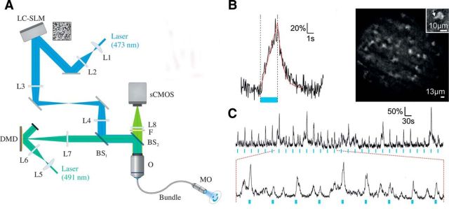
Figure 3.
Patterned photostimulation and functional imaging in freely behaving mice. A, Schematic of the holographic fiberscope composed of two illumination paths: one for photoactivation with CGH including a liquid-crystal spatial light modulator, and a second for fluorescence imaging including a DMD. Backward fluorescence was detected on a scientific complementary metal oxide semiconductor (sCMOS) camera. Both paths were coupled to the sample using a fiber bundle attached to a micro-objective (MO). L, Lens; BS, beam splitter; O, microscope objective. B, Left, Calcium signal triggered by photoactivation (blue line; p = 50 mW/mm2) with a 5 μm holographic spot placed on the soma of a ChR2-expressing cell recorded in a freely behaving mouse coexpressing GCaMP5-G and ChR2 in cerebellar molecular layer interneurons (MLIs). Right, Structure illumination image recorded in a freely behaving mouse and showing MLI somata and a portion of a dendrite (inset). Scale bars: 10 μm. C, Top, The same photoactivation protocol as in A was repeated every 30 s for 15 min (photostimulation power, 50 mW/mm2; imaging power, 0.28 mW/mm2). Bottom, Expansion of the top trace showing that spontaneous activity frequently occurs between evoked transients (adapted from Szabo et al., 2014).

