Figure 1.
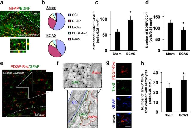
Astrocytes produce BDNF in cerebral white matter in mice. a, Representative images of BDNF (green) and GFAP (astrocyte marker; red) immunostaining in mouse corpus callosum. Z-stack images confirmed that astrocytes produced BDNF. b, Two-month-old male mice were subjected to sham or BCAS surgery and brains were taken out at day 28. Immunohistochemistry using anti-BDNF antibody along with cell-specific marker antibodies (CC1: oligodendrocyte, GFAP: astrocyte, lectin: endothelium, PDGF-R-α: OPC, NeuN: neuron) showed that oligodendrocytes and astrocytes were the major cell types for BDNF production in mouse corpus callosum. The pie charts were generated based on the average of numbers for BDNF+ cells in corpus callosum. n = 5. c, d, Histogram showing the number of BDNF/GFAP-double-positive cells and BDNF/CC1-double-positive cells in mouse corpus callosum. Data are mean ± SD of n = 5. *p < 0.05. e, Representative images of PDGF-R-α (OPC marker; red) and GFAP (astrocyte marker; green) immunostaining in cerebral corpus callosum of 2 months old mice. Scale bar, 100 μm. f, Electron micrograph showing that OPCs attach to astrocytes in 2-month-old rats. Arrowheads indicate PDGF-R-α+ signals. BL, Basal lamina; EC, endothelial cell; Astro, astrocytes. Scale bar, 500 nm. g, Representative images of PDGF-R-α (OPC marker; red), Trk-B (high affinity BDNF receptor; green), and GFAP (astrocyte marker; blue) immunostaining in cerebral corpus callosum of 2-month-old mice. OPCs that expressed Trk-B were located in close proximity to astrocytes. Scale bar, 50 μm. h, Histogram showing the number of Trk-B+ OPCs that showed close contact with astrocytes cells in mouse corpus callosum. Data are mean ± SD of n = 5. *p < 0.05.

