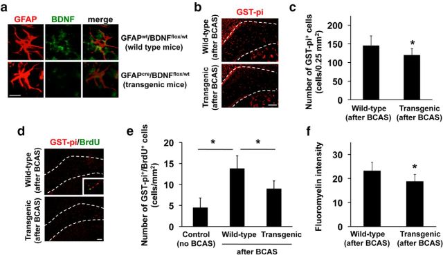
Figure 4.
Astrocyte-derived BDNF supports oligodendrogenesis after white matter injury in mice. a, Representative images of GFAP (astrocyte marker; red) and BDNF (green) immunostaining in corpus callosum of GFAPwt/BDNFfl/wt (wild-type) and GFAPcre/BDNFfl/wt (transgenic) mice. BDNF expression was downregulated in astrocytes in transgenic mice. Scale bar, 20 μm. b, c, Immunostaining of GST-π (oligodendrocyte marker) in mouse corpus callosum. Wild-type (n = 6) and transgenic (n = 5) mice were subjected to prolonged cerebral hypoperfusion by BCAS. At day 28, brains were taken out for immunohistochemistry. Transgenic mice showed lower number of GST-π+ cells. Scale bar, 100 μm. Data are mean ± SD. *p < 0.05. d, e, In vivo OPC maturation assay. Wild-type (n = 6) and transgenic (n = 5) mice were subjected to prolonged cerebral hypoperfusion by BCAS. Mice were treated with BrdU between days 5 and 14. Transgenic mice showed a lower number of GST-π/BrdU-double-positive cells (i.e., newly generated oligodendrocytes) at day 28. Control mice (no BCAS, n = 6) showed the baseline of oligodendrocyte renewal in mouse corpus callosum under normal conditions. Scale bar, 100 μm. Data are mean ± SD. *p < 0.05. f, Fluoromyelin staining showed that myelin density in corpus callosum region at day 28 in GFAPcre/BDNFfl/wt mice was lower than that in GFAPwt/BDNFfl/wt mice. Data are mean ± SD. *p < 0.05.

