Figure 2.
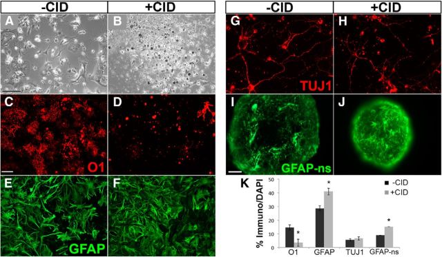
CID treatment of neonatal MBP-iCP9 cultures induces cell death selectively in O1+ oligodendrocytes. A, B, Bright-field microscopy indicates extensive cell death 24 hours after treatment with 10 μm CID. C, D, Labeling with the mAb O1 demonstrates significant and selective depletion of oligodendrocytes in CID-treated cultures. By contrast, neither GFAP+ astrocytes (E, F) nor Tuj1+ neurons (G, H) were reduced by CID treatment, demonstrating that CID is specific for oligodendrocyte death. The number of GFAP+ cells increased significantly in dissociated cultures as well as in CID-treated neurosphere preparations (I, J). K, Summary histogram of cell counts of oligodendrocytes (O1), astrocytes (GFAP), neurons (Tuj1), and neurosphere-derived astrocytes (GFAP-ns) from MBP-iCP9 cultures with vehicle or CID treatment. n = 3 coverslips per condition per cell marker repeated in cultures from 2 or 3 different mice. *p < 0.05. Error bars indicate SEM. Phase image scale bar, 100 μm; white scale bar, 30 μm, applies to all fluorescent images, except 50 μm scale bar in I.

