
Figure 5.
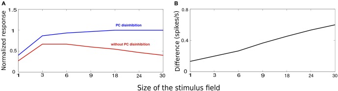
VIP-mediated disinhibition. The stimuli chose for this experiement is similar to Figure 2A with the size of the stimulus controlled by the number of activated minicolumns. (A) Average minicolumnar response during absence (red) and presence (blue) of VIP-mediated disinhibition normalized to peak response. (B) The vertical axis denotes the difference between the responses in absence of presence of VIP-mediated disinhibition. The effect of disinhibition grows with the stimulus size.