
Fig. 2.
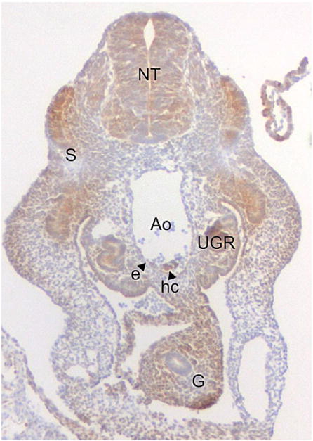
Cellular hypoxia in the E10 mouse embryo. A transverse section through 33 somite pair embryo stained for the hypoxia indicator Pimonidazole (hypoxyprobe) is shown. Hypoxic cells (brown) are found in the neural tube (NT), gut (G), somites (S), aorta (Ao) and urogenital ridges (UGR). Arrow heads indicate hypoxic aortic endothelial cells (e) and hematopoietic cluster cells (hc).