
Fig.3.
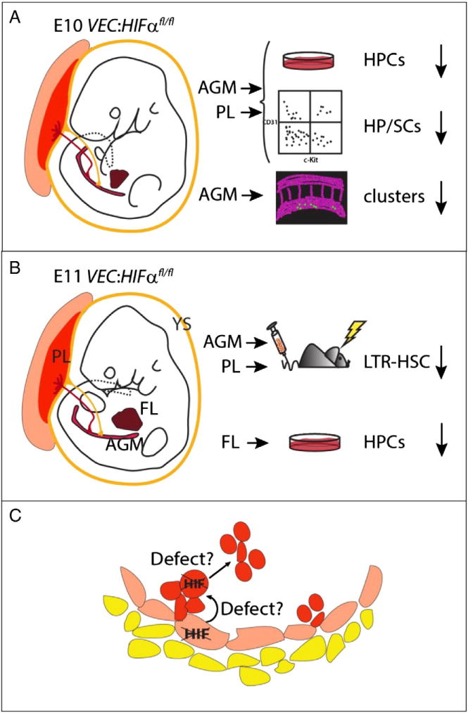
Hematopoiesis defects in VEC-Cre/+:HIF1αfl/fl embryos.A) E10 and B) E11 mouse VEC-Cre/+:HIF1αfl/fl embryos are depicted at the time when the first hematopoietic progenitor and stem cells (HPSCs) are generated in the aorta. Tissues where hematopoietic cells appear in the embryo are shown: The extraembryonic yolk sac (YS) and placenta (PL), the intraembryonic aorta–gonad–mesonephros (AGM) and fetal liver (FL). The results of CFU-C progenitor (HPC), phenotypic (FACS) analysis for HP/SC, 3-D imaging for aortic hematopoietic clusters and long-term in vivo reconstitution assays are shown. C) Schematic close-up of the ventral wall of the aorta showing hematopoietic cluster emergence from hemogenic endothelium in VEC-Cre/+:HIF1αfl/fl embryos. Defects in HPSCs detected by different hematopoietic assays are proposed in the hemogenic endothelium and/or in hematopoietic cell emergence/expansion/maintenance [60].