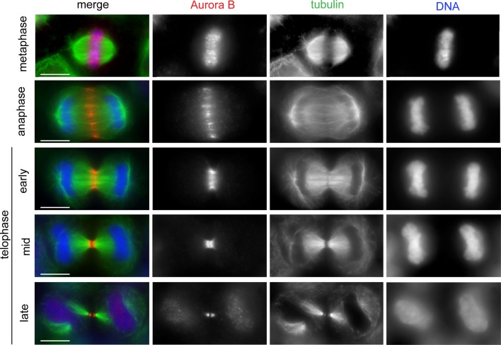
Figure 1.
The CPC shows dynamic localization during mitosis and cytokinesis. HeLa cells were fixed and stained to reveal Aurora B (red), tubulin (green), and DNA (blue). The CPC (here represented by Aurora B) translocated from the mitotic chromosomes to the central spindle early in anaphase. In early telophase, the CPC accumulated at the midbody arms. Scale bars: 10 μm.

