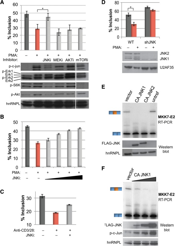
Figure 3.
JNK activity is necessary and sufficient for activation-induced MKK7-E2 skipping. (A, top) Average percent MKK7-E2 inclusion (n = 3) in Jurkat T cells pretreated with the following inhibitors prior to PMA treatment: 50 μM JNKi (SP600125), 20 μM MEKi (U0126), 1 μM Akti (triciribine), and 100 nM mTORi (rapamycin). (Bottom) Western blot analysis of downstream substrates phospo-c-Jun (JNK), phospho-Erk (MEK), and phospho-S6K (mTOR); the Akt-activating autophosphorylation site Ser473 (Akt); and hnRNPL as a loading control. Protein samples were harvested 2 h after PMA, while RNA was harvested after 48 h. Note that the samples from mTOR inhibition were run on the same gels as the other samples but with a spacer lane that has been removed. (B) Average MKK7-E2 inclusion (n = 3) with 12.5, 25, 50, and 100 μM JNK inhibitor SP600125 prior to PMA treatment (48 h). (C) Average MKK7-E2 percent inclusion (n = 2) of primary human CD4+ T cells treated with JNK inhibitor SP600125 and/or anti-CD3/CD28 (48 h). See also Supplemental Figure S2B. (D) Average percent MKK7-E2 inclusion (top) and Western analysis (bottom) in two independent Jurkat T lines depleted of JNK by an shRNA grown in the absence or presence of PMA (48 h). (E) Representative (n = 3) RT–PCR gel of MKK7-E2 inclusion (top) and the corresponding Western analysis of Flag-CAJNK expression (bottom) from HEK293 cells transfected with vector control or constitutively active JNK1 and JNK2 (CAJNK1 or CAJNK2). (F) Same as in E but with 0.01, 0.1, and 1 μg of CAJNK1. Phospho-c-Jun, as a marker for CA-JNK activity, is also shown. Error bars represent standard deviation. (*) P < 0.005, Student's t-test. See also Supplemental Figure S2.

