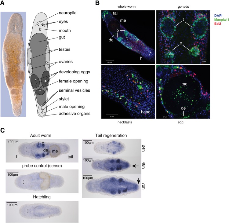
FIGURE 3.
Expression patterns of Macpiwi1 and Macpiwi2 in M. lignano. (A) Interference contract image and diagrammatic representation of an adult worm. (B) Immunofluorescence labeling showing localization of Macpiwi1 (green) in adult worms. Dividing cells are labeled with EdU (red). (h) Head, (t) testis, (o) ovary, (de) developing egg, (me) mature egg. Stars denote eyes. Arrow points to nonspecific staining by the secondary antibody. (C) Localization of Macpiwi2 mRNA by whole-mount in situ hybridization in adult worms, hatchlings, and during regeneration induced by posterior amputation. Sense riboprobe was used as negative control. Arrows point to blastemas during regeneration. (t) Testis, (o) ovary, (de) developing egg, (me) mature egg.

