
FIGURE 3.
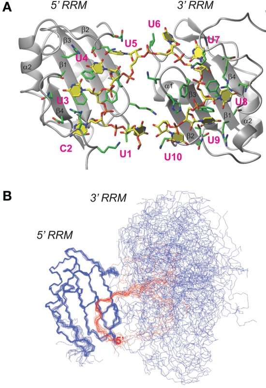
Structural model of the RRM dimer. (A) Representative structure of the double-RRM/UC(U)8 model obtained by torsion angle dynamics calculation. Protein is shown in ribbon and RNA in stick representation. Nucleotides are numbered in magenta. (B) Bundle of 20 structures with the lowest CYANA target function. Protein backbone shown in blue and RNA in red. The ensemble is aligned to the backbone coordinates of the 5′ end-binding RRM in order to show the inter-RRM flexibility. Backbone pairwise RMSD of the 5′ end UC(U)3 RNA–RRM complex is 0.56 Å, that of the 3′ end (U)5 RNA–RRM complex is 0.52 Å, and the RMSD of the 2:1 RRM: UC(U)8 complex is 8.16 Å.