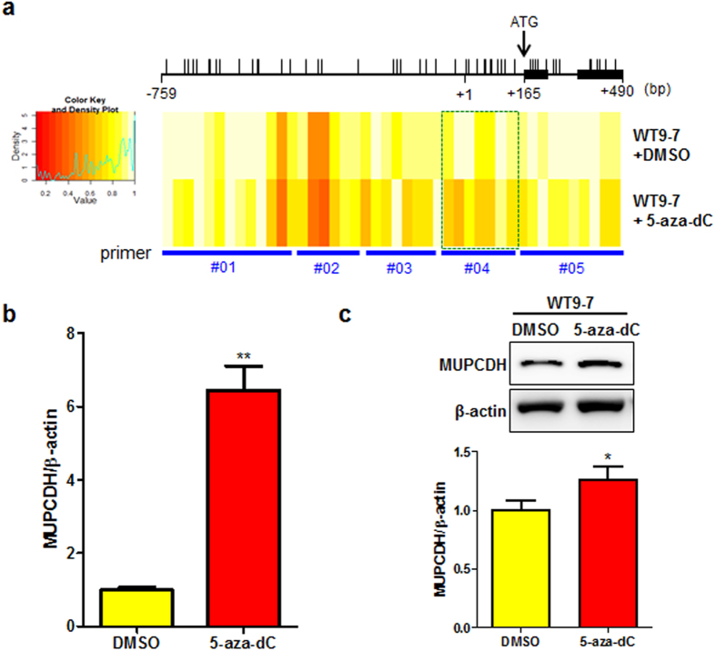
Figure 3. Restoration of the MUPCDH expression level by treatment with 5-aza-2′-deoxycytidine (5-aza-dC) in ADPKD cyst-lining epithelial cells.
(a) The altered DNA methylation pattern in the MUPCDH promoter region was confirmed using the EpiTYPER® assay. As shown in the EpiTYPER® heat map, methylated CpG sites (yellow) were changed to unmethylated CpG sites (red) after treatment of WT9-7 cystic epithelial cells with 5-aza-dC (5 μM, 72 h). Green box indicate CpG sites within −543 and +228, which was more sensitive to 5-aza-dC, compared to those in other regions. The translation start ATG is indicated by a black arrow. P < 0.05. (b) Altered mRNA and protein expression levels upon treatment of WT9-7 cells with 5-aza-dC were confirmed by real-time quantitative reverse transcription polymerase chain reaction (qRT-PCR) analysis, and (c) western blot analysis. The band density was measured using the MultiGauge software. The height of each bar represents the mean, and the error bars indicate ± SD. β-Actin was used as an internal control in real-time qRT-PCR and western blot analyses. Each experiment was performed in triplicate. *P < 0.05; **P < 0.001.

