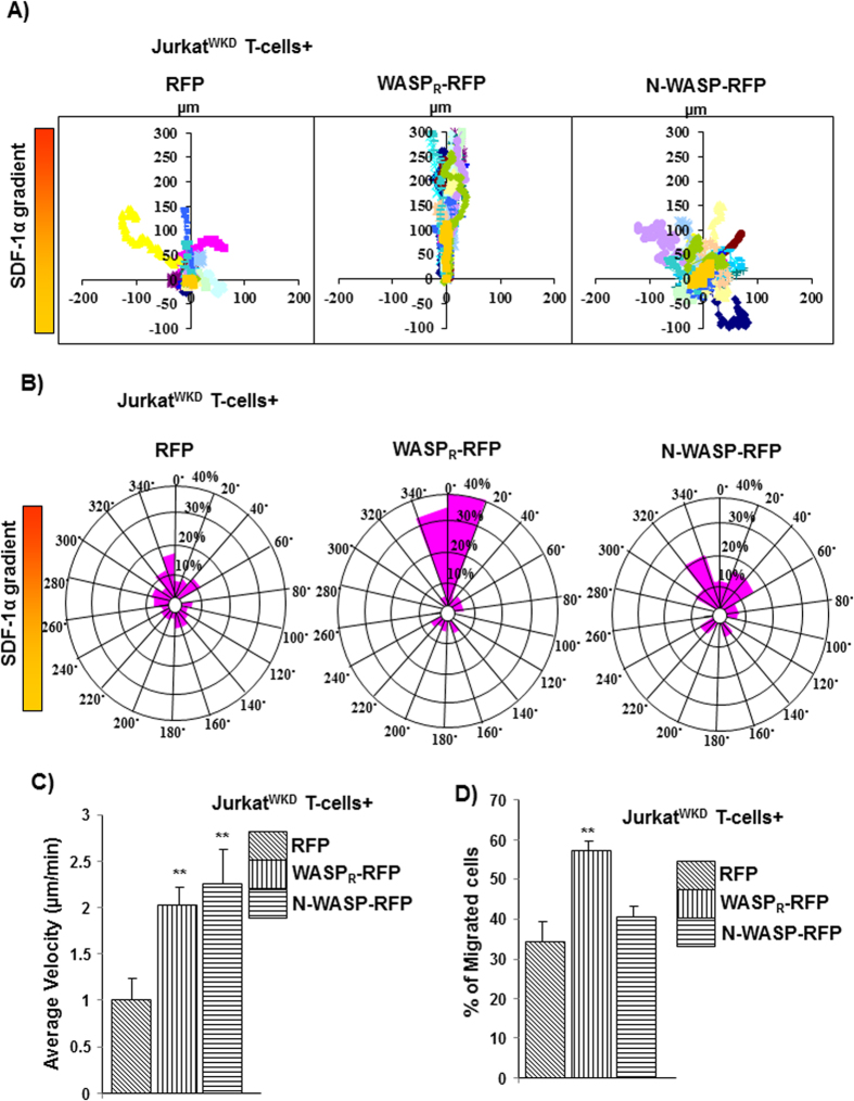
Figure 2. N-WASP expression does not rescue the WASP deficiency in Jurkat T-cells chemotaxis.
(A) Vector plots representing migration path of 20 randomly selected JurkatWKD T-cells expressing (1) RFP, (2) WASPR-RFP, (3) N-WASP-RFP in Dunn chamber assay exposed to a gradient of chemokine SDF-1α (maximum at top). The intersection point of X and Y axis was taken as starting point of each cell. (B) Overall directionality of migration (final position of cell in each 20° sector). (C) Migration velocity of total 60 randomly selected cells of cell type as in panel A. **P < 0.01 compared to RFP expressing JurkatWKD T-cells. (D) Transwell migration of JurkatWKD T-cells expressing (1) RFP, (2) WASPR-RFP, (3) N-WASP-RFP represent as percentage of cells migrated. **P < 0.01 compared to RFP expressing JurkatWKD T-cells.

