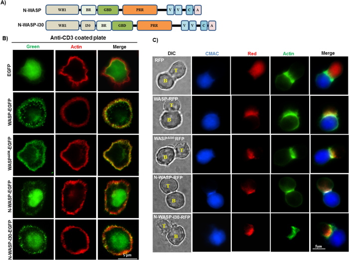
Figure 5. I30 region of WASP recruit N-WASP-I30 to punctate structures at cell cortex.
(A) WASP-I30 region was inserted in N-WASP between WH1 domain and basic region of N-WASP. (B) Jurkat T-cells expressing EGFP tagged WASPR, WASPRΔI30, N-WASP and N-WASP-I30 were stimulated on anti-CD3 (OKT3)-coated coverslip for 5 min and stained with Alexa fluor 594 phalloidin. The experiment has been repeated three times and 25 cells were analyzed each time. (C) Jurkat T-cells expressing WASPR-RFP, WASPRΔI30-RFP, N-WASP-RFP and N-WASP-I30-RFP were allowed to form conjugate with Raji B cell initially pulsed with SEE toxin. The conjugates between T: B cells were detected by Alexa fluor 488 phalloidin. Bar = 5 μm.

