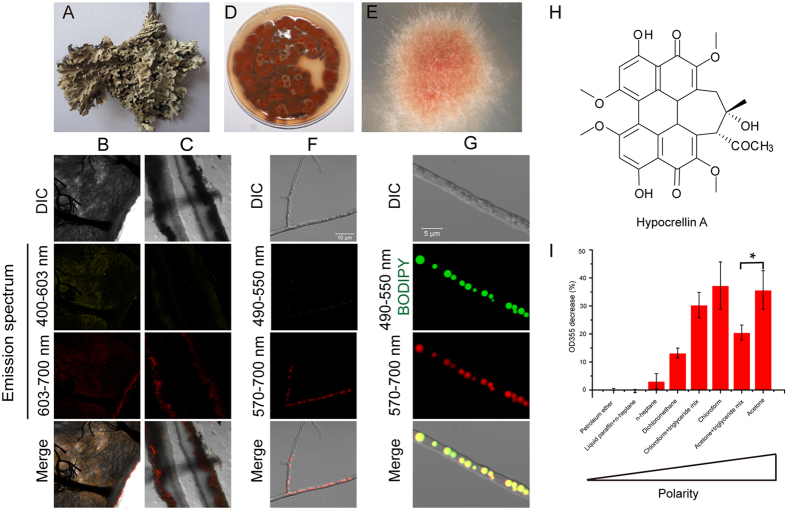
Figure 1. Trapping phototoxins in lipid droplets probably serves as a resistance mechanism in an endolichenic fungus.
(A) An image of the thallus of lichen, Heterodermia obscurata (Nyl.) Trevis. (B,C) CLSM observation of a thallus cut transversely (B) or longitudinally (C). (D,E) Photomicrographs of the isolated endolichenic fungus colony. (F) Filaments of isolated fungi, observed by CLSM. The fluorescent substances produced by filaments were excited by 488 nm and 555 nm lasers and observed at the two indicated emission spectrums. The fluorescent substances within the filaments displayed red fluorescence in the emission spectrum of 570–700 nm rather than 490–550 nm. (G) Intracellular distribution of fluorescent substances. The filaments were stained with BODIPY, a dye that is specific for LDs, and observed by CLSM using the same conditions described in panel (F). Emission spectrums of 490–550 nm and 570–700 nm were used to detect BODIPY staining and fluorescent agents, respectively. (H) The chemical structure of HA. (I) Detection of singlet oxygen molecules produced by HA under light irradiation in different solvents. Data represent mean values ± SD from three independent replicates.

