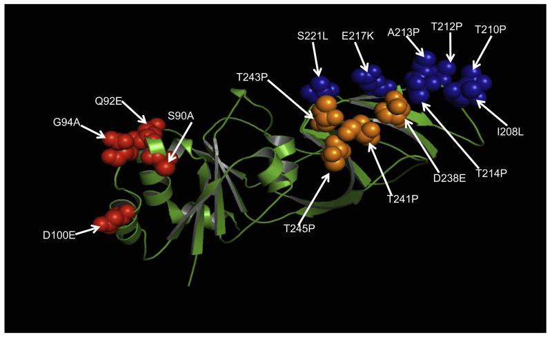
Fig. 3.
Substitutions in strain RVA/Human-wt/CMR/6788/1999/G9P[8] highlighted on the crystal structure of RRV VP7 protein (3fmg). The molecule is colored in green. Residues corresponding to previously describe major antigenic sites A, C and F are indicated in red, blue and orange spheres, respectively. Arrows indicate substitutions at amino acid positions. (For interpretation of the references to colour in this figure legend, the reader is referred to the web version of this article.)

