Figure 2.

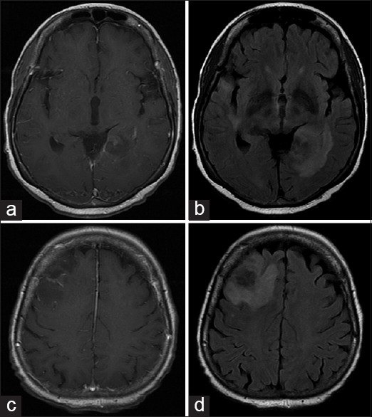
Magnetic resonance imaging brain T1 with contrast (a and c) and fluid-attenuated inversion recovery (b and d) demonstrates multifocal, patchy enhancing lesions at right frontal and left periatrial regions

Magnetic resonance imaging brain T1 with contrast (a and c) and fluid-attenuated inversion recovery (b and d) demonstrates multifocal, patchy enhancing lesions at right frontal and left periatrial regions