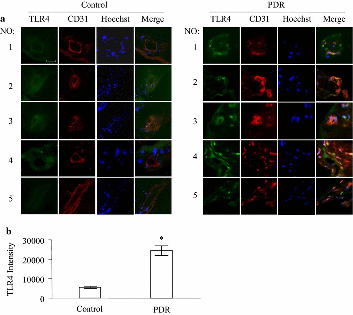
Fig. 1.

Expression of TLR4 in PDR fibrovascular membrane. Immunofluorescence staining of TLR4 (green) and CD31 (red) was performed in normal human retinas, fibrovascular membranes from PDR patients. Results show five fibrovascular membranes from patients with PDR and five normal human eye ball controls (a). TLR4 immunofluorescence relative intensity in retinal sections was quantified and displayed (b). *P < 0.05 compared to the control group. Scale bar 20 μm
