Figure 1.
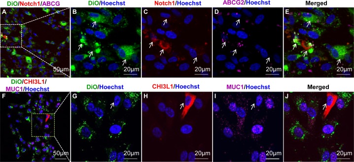
TMSCs are slow-cycling in vitro. Cultured TMSCs were labeled with lipophilic dye DiO (green) and, after two passages, incubated with Hoechst 33342 for 1 hour, followed by staining with Notch1 (red) and ABCG2 (purple, [A–E]) or CHI3L1 (red) and MUC1 (purple, [F–J]). Arrows in (B–E) point to DiO-retaining cells; arrows in (G–J) point to DiO-negative CHI3L1 expression cells. DAPI stains nuclei as blue.

