Figure 7.
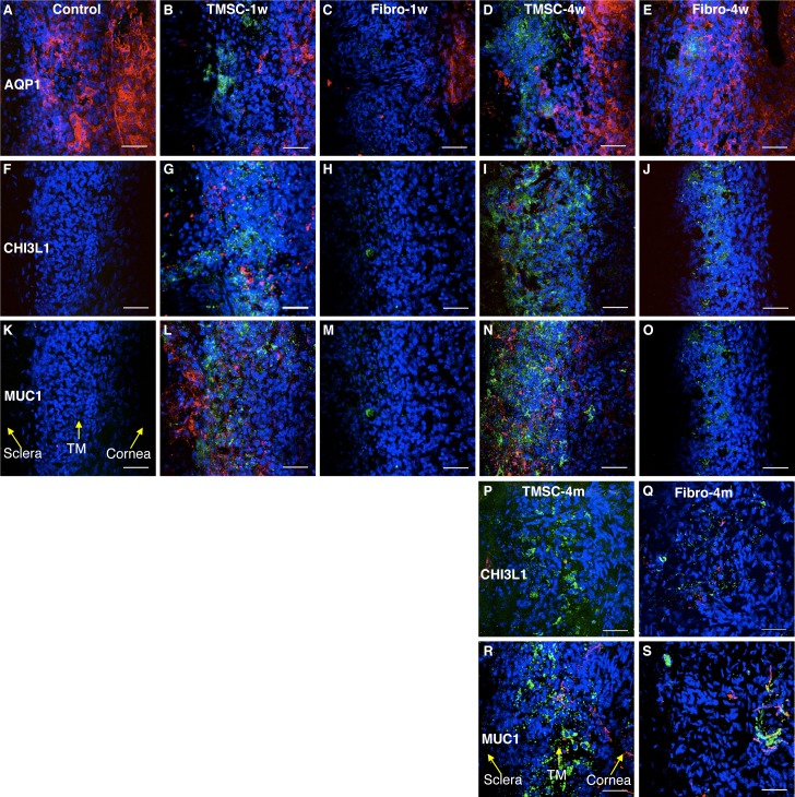
Human TMSCs home to TM region after mouse anterior chamber injection. DiO-labeled human TMSCs or fibroblasts were injected into normal mouse anterior chamber for 1 week, 4 weeks, and 4 months. Wholemount stain was performed to detect the green-injected cells and the expression of AQP1 (red; [A–E]) and human specific antigens CHI3L1 (red; [F–J, P, Q]) and MUC1 (red; [K–O, R, S]) on host TM region. (A, F, K) show staining of the antibodies on normal mouse tissue without cell injection serving as controls. (B, G, L) show staining on TMSC-injected tissue at 1 week. (C, H, M) show staining on fibroblast-injected tissue at 1 week. (D, I, N) show staining on TMSC-injected tissue at 4 weeks. (E, J, O) show staining on fibroblast-injected tissue at 4 weeks. (P, R) show staining on TMSC-injected tissue at 4 months. (Q, S) show staining on fibroblast-injected tissue at 4 months. DAPI stains nuclei as blue. The orientation of the tissue is indicated by yellow arrows in (K, R). Scale bars: 50 μm.

