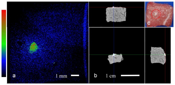
Fig. 4.

Bone on tissue imaging: a. Top view Raman computed image of the tissue sample field of view is 1 cm. b. microCT projections of the sample, top and perpendicular side views. c. Top view color image with bone fragment encircled in red, bone size is ~1 mm.
